Electroretinography in Dogs: Measuring Retinal Function Before Clinical Signs Appear
The electroretinogram (ERG) is the gold standard for detecting retinal dysfunction before structural changes become visible. Understanding what ERG measures, how the procedure works, and when it is indicated in PRA diagnosis.
The ophthalmoscope reveals what has already been lost. When I identify the characteristic changes of PRA on funduscopic examination — the increased tapetal reflectivity, the attenuated vessels, the pallid optic disc — I am documenting damage that is already substantial. Rod photoreceptors must degenerate significantly before these structural changes become visible.
Electroretinography allows me to detect the problem earlier. The ERG measures the electrical response of the retina to controlled light stimuli, providing an objective, quantitative assessment of photoreceptor function independent of the structural changes visible on ophthalmoscopy. In breeds at risk for PRA, ERG can confirm retinal dysfunction months to years before ophthalmoscopic changes appear.
What the Electroretinogram Measures
When light strikes a photoreceptor, it triggers a cascade of molecular events that ultimately generate an electrical signal. This signal propagates through the retinal layers, generating measurable electrical potentials at the surface of the eye. The ERG records these potentials using electrodes placed on the corneal surface.
The resulting waveform has characteristic components. The a-wave represents primarily photoreceptor activity — its amplitude reflects the functional mass of rod and cone cells. The b-wave arises from bipolar cells and Müller glia in the inner retina, reflecting the transmission of signals from photoreceptors to deeper retinal layers. Oscillatory potentials superimposed on the b-wave reflect amacrine cell activity in the inner retina.
Scotopic a-wave: Rod photoreceptor function (dim-light stimuli)
Scotopic b-wave: Rod pathway transmission
Photopic b-wave: Cone photoreceptor function (bright-light stimuli)
Implicit times: Speed of response — delays indicate dysfunction
Flat ERG: No measurable response — complete photoreceptor failure
In early PRA, the scotopic responses diminish first — reflecting rod degeneration — while photopic (cone) responses remain near normal. This electroretinographic profile mirrors the clinical presentation described in our guide on how PRA progresses clinically: night blindness preceding day vision loss.
The ERG Procedure in Practice
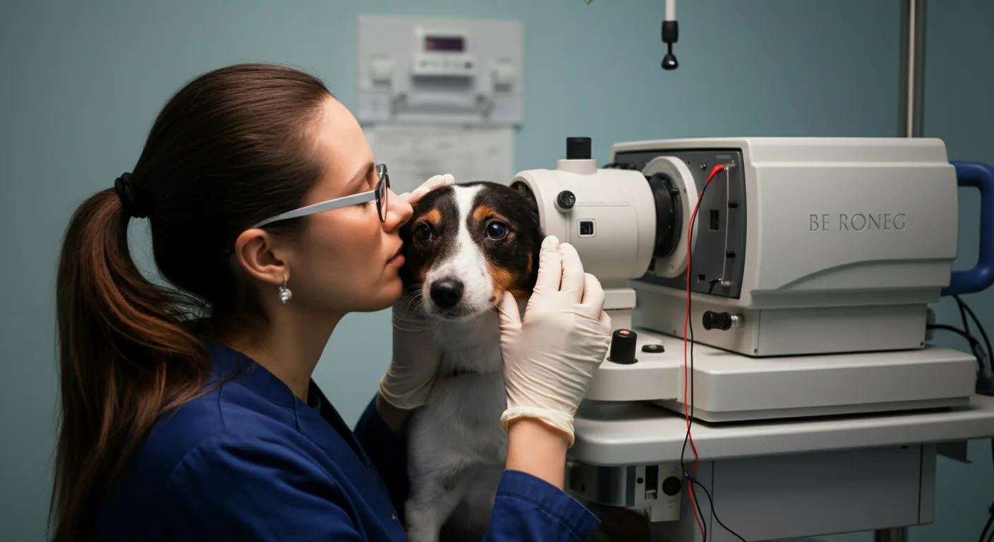
Performing an ERG in dogs requires sedation or general anesthesia. The dark-adapted state requires at least 20–30 minutes of dark adaptation before recording, making the procedure time-intensive. Corneal electrodes, typically contact lens-style or small fiber types, are placed on the corneal surface with a conductive medium. Reference and ground electrodes are placed at standard locations on the head.
A complete diagnostic ERG protocol includes:
- Dark-adapted (scotopic) testing: Multiple flash intensities delivered in dark conditions to characterize rod system function
- Light-adapted (photopic) testing: Recording after ten minutes of bright background illumination to isolate cone system responses
- Flicker ERG: High-frequency stimulus to assess cone temporal resolution
The entire recording session typically takes 45–90 minutes including dark adaptation. Results are compared against age-matched normal values established for the breed or species being tested. Amplitude reductions and implicit time changes indicate retinal dysfunction even when ophthalmoscopic examination appears normal.
When Is ERG Indicated in PRA Assessment?
ERG serves several roles in PRA diagnosis and management:
Pre-Breeding Screening in High-Risk Breeds
For breeds where DNA testing is unavailable or incomplete, ERG provides functional evidence of retinal health. Annual examination by a board-certified ophthalmologist identifies dogs developing ERG changes before clinical signs appear, allowing breeders to remove them from breeding programs before affected offspring are produced. This approach was the cornerstone of PRA management before molecular tests became available.
Distinguishing PRA from Other Retinal Conditions
Several retinal conditions mimic PRA ophthalmoscopically. Sudden acquired retinal degeneration syndrome (SARDS), inflammatory retinopathies, and nutritional deficiencies can produce fundus changes superficially similar to PRA. ERG helps distinguish these conditions: SARDS produces an acutely flat ERG even when the fundus appears relatively normal initially; inflammatory conditions may show selective inner retinal dysfunction rather than the outer retinal pattern of PRA.
Monitoring Progression
Serial ERG recordings quantify the rate of photoreceptor loss over time. This information helps counsel owners about expected progression timeline and, increasingly, identifies candidates for gene therapy interventions where sufficient photoreceptor viability must remain for treatment to succeed.
Confirming Diagnosis Before Breeding Decisions
When DNA testing yields unexpected results — an apparent clear dog showing clinical signs — ERG provides independent functional confirmation. Conversely, a carrier dog showing equivocal ophthalmoscopic changes can be evaluated with ERG to determine whether genuine retinal dysfunction is present.
ERG vs. DNA Testing
ERG and DNA testing provide complementary information and serve different roles. DNA testing identifies genetic status — clear, carrier, or affected — before any clinical or functional changes develop. It can be performed at any age, including before weaning if necessary. For breeds where the causative mutation is known, DNA testing is the preferred primary screening tool because it identifies carriers who will never develop disease themselves but can pass it to offspring.
ERG detects functional change in the retina but cannot identify carriers. A carrier dog with normal retinal function will have a completely normal ERG — which is expected, since carriers do not develop disease. ERG also cannot distinguish between different PRA mutations based on waveform characteristics alone.
Use DNA testing when the causative mutation is known for your breed. It identifies carriers, not just affected dogs, and guides breeding decisions most effectively.
Use ERG when no DNA test is available for your breed's PRA form, when distinguishing PRA from other retinal conditions, when monitoring progression in affected dogs, or when DNA results are unexpected and functional confirmation is needed.
ERG in Research and Therapeutic Trials
ERG is the primary efficacy endpoint in virtually all PRA gene therapy research. Before and after recordings in the same eyes demonstrate whether therapeutic gene delivery has restored photoreceptor function. The amplitude improvements seen in early gene therapy trials — such as the landmark RPE65 work in Briards that led to the development of Luxturna — were documented through ERG recordings showing restoration of scotopic responses in treated eyes compared to untreated controls.
For owners following research progress, ERG data in published trial results provides objective evidence of treatment efficacy independent of behavioral assessments. A dog navigating an obstacle course more confidently after treatment is compelling, but the ERG demonstrating restored photoreceptor responses is the quantitative foundation of the result.
Access to ERG testing requires a veterinary ophthalmologist or veterinary neurologist with appropriate equipment and expertise. Board-certified veterinary ophthalmologists can be located through the American College of Veterinary Ophthalmologists (ACVO) or European College of Veterinary Ophthalmologists (ECVO) directories. For owners seeking diagnostic confirmation of suspected PRA, understanding what a full retinal examination involves helps prepare for the consultation. Combined with appropriate DNA testing for breed-relevant mutations, ERG provides the most complete picture available of a dog's retinal health status.
Dr. Amanda Foster, Veterinary Ophthalmologist