Ophthalmoscopy and Retinal Examination in Dogs: What Veterinary Ophthalmologists Look For
A detailed look at how veterinary ophthalmologists examine the canine retina, what changes indicate PRA at different stages, and why regular eye certification matters for breeding dogs.
The fundus of the canine eye is a window into some of the most important genetic health information available. When I perform an ophthalmoscopic examination, I am not merely checking for acute disease. I am reading a visual record of the dog's retinal health, looking for the subtle early changes that herald conditions like Progressive Retinal Atrophy months or years before they become obvious to owners.
Understanding what this examination involves — what we are looking for and what changes mean — helps breeders and owners make sense of eye certification reports and understand why annual examinations add value beyond the baseline DNA test.
The Normal Canine Fundus
The canine fundus (back of the eye) has several distinctive features that differentiate it from the human fundus and provide the baseline against which changes are assessed:
The tapetum lucidum is a specialized reflective layer present in the dorsal fundus of dogs, responsible for the characteristic eye-shine seen in photographs or headlights. The tapetum appears as a brightly reflective zone in shades of green, gold, or blue depending on the individual dog and breed. Its condition and reflectivity are important diagnostic indicators.
The non-tapetal fundus occupies the ventral portion of the fundus and appears dark brown or slate, with pigmentation influenced by coat and eye color. The distribution and uniformity of this pigmentation provides additional diagnostic information.
The optic disc (optic nerve head) appears as a roughly triangular or circular pale structure, typically in the non-tapetal region in most breeds. Its color, size, and sharpness of borders are assessed.
The retinal vasculature consists of arterioles and venules radiating from the optic disc. The caliber, color, and regularity of these vessels provide important clues to retinal health.
Ophthalmoscopic Changes in PRA by Stage
The characteristic changes of PRA follow a predictable sequence as the disease progresses. Recognizing these stages helps interpret eye certification reports and understand where a dog is in the disease process. The detailed progression is covered in our overview of how PRA advances from night blindness to complete vision loss.
Early Changes
The earliest ophthalmoscopic change in most PRA forms is increased tapetal reflectivity. The tapetum appears brighter than expected for the dog's age, with a more uniform, mirror-like quality replacing the normal slightly granular texture. This change reflects early thinning of the overlying photoreceptor layer, allowing more light to reach and reflect from the tapetum.
Concurrent with or shortly after the reflectivity change, the retinal arterioles begin to show subtle attenuation. Normal arterioles appear as bright, clearly defined red vessels. Early PRA produces slight reduction in vessel caliber and a subtle change in vessel reflex. These changes may require comparison with normal breed-matched photographs to appreciate.
Moderate Changes
As PRA advances, the tapetal reflectivity becomes more pronounced and may extend into regions that were previously less reflective. The tapetum may show an abnormally uniform, bright green or gold appearance. Vascular attenuation becomes more obvious, with arterioles appearing thin and wire-like compared to normal.
The optic disc begins to show pallor — a fading of the normal pink-orange color toward white or grey. This reflects degeneration of the retinal nerve fiber layer as photoreceptors and their associated neurons degenerate. Disc pallor is a relatively late finding but confirms significant retinal damage.
Advanced Changes
In advanced PRA, the fundus presents a characteristic appearance: hyperreflective tapetum, severely attenuated or nearly invisible vessels, and a pale, waxy optic disc. The non-tapetal fundus may show changes in pigmentation as retinal pigment epithelium cells are affected.
At this stage, lens changes may also be visible. Secondary posterior subcapsular cataracts develop in many PRA-affected dogs as degenerating retinal tissue releases abnormal metabolites. These cataracts appear as an opacity at the posterior pole of the lens, visible on slit-lamp examination.
Examination Techniques
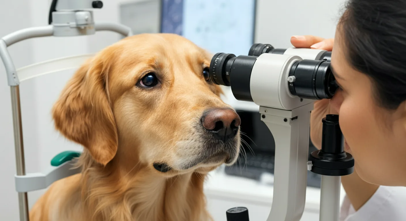
Veterinary ophthalmologists use several instruments and techniques to examine the canine fundus:
Indirect Ophthalmoscopy
The most common clinical technique involves a headband-mounted light source and a condensing lens held before the eye. This method provides a wide-field, stereoscopic view of the fundus, allowing the examiner to survey the entire retina systematically. Most veterinary eye certification examinations use indirect ophthalmoscopy as the primary technique.
Direct Ophthalmoscopy
A handheld direct ophthalmoscope provides a magnified, upright view of a smaller fundus area. This technique offers higher magnification for detailed assessment of specific regions, such as the optic disc or individual vascular changes, but has a narrower field of view.
Slit-Lamp Biomicroscopy with Condensing Lens
Combining a slit-lamp microscope with a handheld condensing lens provides an excellent fundus view with variable magnification and illumination. This technique is particularly useful for detailed examination of the vitreous and posterior segment. It is also the standard technique for anterior segment examination, including assessment of the lens for secondary cataracts.
Pupil Dilation for Examination
Thorough fundus examination requires pharmacological pupil dilation. Mydriatic drops — typically tropicamide in veterinary patients — are applied to each eye before examination. Full dilation takes 15–20 minutes and persists for several hours. Dogs should not be expected to navigate in bright light immediately after dilation, and drivers bringing animals for certification should plan accordingly.
Some breeds with narrow iridocorneal angles may be at risk for post-dilation secondary glaucoma. This is uncommon in most breeds but is considered in susceptible individuals before dilation.
Eye Certification Programs
Annual eye certification through veterinary ophthalmologists provides a systematic, breed-aware assessment of retinal health. Certification schemes exist in most countries with significant canine breeding populations:
- CERF/OFA Eye Certification (USA): The Orthopedic Foundation for Animals maintains eye examination records. Examinations must be performed by board-certified ACVO diplomates.
- BVA/KC Eye Scheme (UK): The British Veterinary Association coordinates examinations by specially trained veterinary surgeons.
- ECVO Eye Scheme (Europe): The European College of Veterinary Ophthalmologists coordinates breed-appropriate examination standards across European countries.
Certification provides documentation of clinical normality at the time of examination but cannot rule out early PRA in a dog that will develop clinical signs later. This is why DNA testing, which identifies genetic status independent of clinical stage, remains the more powerful tool for breeds with known mutations. Our overview of breed health registries and PRA documentation programs explains how to access certification records and contribute test results for breeding partners.
What to Expect at an Eye Certification Appointment
For breeders unfamiliar with the process, a standard eye certification appointment proceeds as follows: brief case history and observation, anterior segment examination with slit-lamp, pupil dilation and 15-minute wait, indirect ophthalmoscopy of both fundi, and issuing of a certification form documenting findings.
The examiner will note any abnormalities, classify them according to breed-specific criteria (some changes are breed-normal variants not related to disease), and indicate whether the dog passes certification. Results are submitted to the relevant registry and become part of the dog's permanent health record.
When combined with DNA testing for known PRA mutations and, where indicated, electroretinography for functional assessment, annual ophthalmoscopic examination provides the most complete picture available of a breeding dog's retinal health. For breeders committed to evidence-based health programs, this combination represents the current standard of care.
Dr. Amanda Foster, Veterinary Ophthalmologist